
An introduction to building the blocks of the iToBoS solution: from fundamentals to camera testing. As we are designing the whole imaging system from scratch, many parameters regarding the imaging system of the total body scanner needed to be specified.
Foremost the fundamentals
The working horse of the body scanning system is an imaging camera and its associated optical components. The cameras are responsible for taking the images for the medical professionals as well as the input for the artificial intelligence algorithms. Without an iota of doubt, the functionality and end quality of the scanning is de- pendent on the camera characteristics.
For the top-notch cameras, parameters like frame rate, field of view, resolution, and pixel size are decisive factors to be chosen for the intelligent total body scanner. With that in mind we contacted several camera distributors in Germany, who gave their expertise to the camera selection process. We decided upon goal-specifications that the imaging system should deliver. Important specifications of the imaging system are for example the image resolution and a natural color perception. Beyond that the acquisition speed plays a huge role. It is important for the camera to be fast to reduce the time that the patient has to spend within the scanner. Beyond that, it is important for the medicals to have very natural colors. Furthermore, many other components, as for example the illumination, depend upon the choices in the imaging system as well.
With this in mind we started the camera testing starting with the resolution that we would like to explain in more detail in this blogpost. This was done on artificial targets as well as on human skin samples.
Testing the cameras
The diffraction limited optical resolution is a parameter that describes to what detail an object can be shown. For example, to resolve a human hair which is µm one needs a resolution of exactly that width to ensure that the hair is visible. For the intelligent total body scanner, a target resolution has been called out taking the sizes of the bio tissue in melanoma into account.
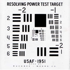
USAF target. (www.optowiki.info/faq/how-to-read-an-usaf1951-target)
From an optics perspective the resolution can be measured with a USAF-target. This measurement method has been developed by the US army in the 1950s. It consists of groups of parallel lines with different distances to each other. The higher the group number the smaller is the distance between the parallel lines and the higher is the resolution.
In detail the resolution measurement is done by taking a photograph with the imaging system under test. The acquired image is analyzed further with image analysis software. Every group will be studied by eye whether the lines are still distinguishable. If so, the group is resolved.
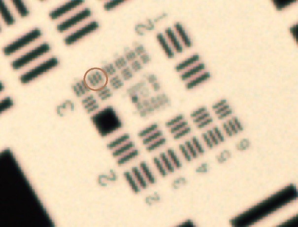
USAF target. (www.optowiki.info/faq/how-to-read-an-usaf1951-target)
A simple calculation will then give the resolution in Micrometers. These resolutions measurements need to be done for different measurements to find the optimal working distance for the final intelligent total body scanner.
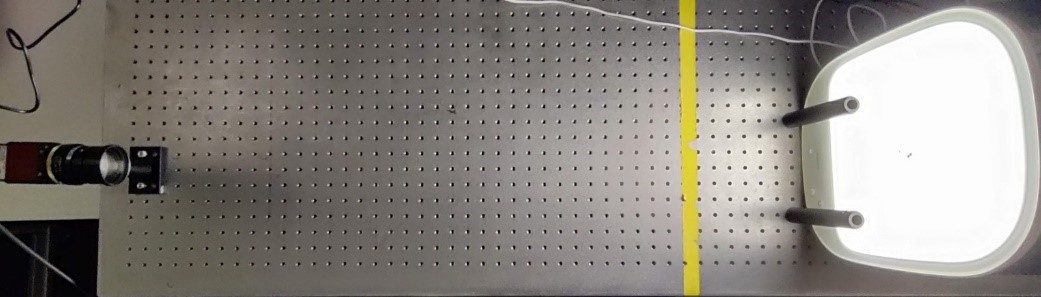
Setup for resolution measurements.