
The practice of pathology has undergone a foundational change over the last decade, largely thanks to the widespread adoption of whole slide imaging (WSI).
The advances in the capture and management of the whole slide images led to the rapid increase of the data volume. This, in turn, has forced pathologists to embrace tech solutions that facilitate more efficient data processing.
Computer vision, in particular, assists pathologists with such tasks as:
- Image analysis and interpretation
- Early disease detection and prevention
- Sample tissue inspection and anomaly detection
- Matching pathology types with historical cases
And that’s just the tip of the iceberg.
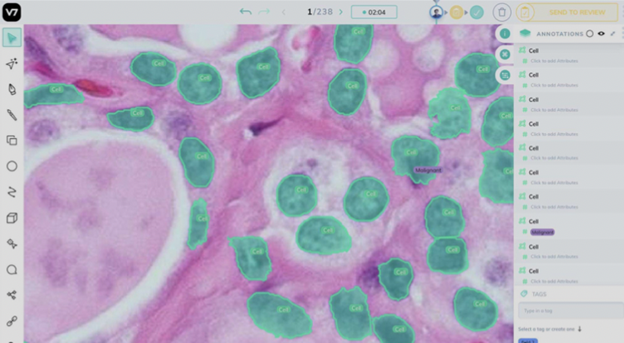
Here’s an example of the cell annotation in a microscopic image that can be used to train AI models for disease detection, such as cancer:
To understand how promising the employment of computer vision is for the future of digital pathology, you only need to glance at the Camelyon Grand Challenge 2016 results.
The program aimed to “evaluate new algorithms for the automated detection of cancer metastases on gigapixel pathology images.” The AI algorithm has achieved a stunning 94.2% sensitivity in tumor detection rate, while human pathologists reached only 73.2%.
The future of computer vision in digital pathology might seem bright and optimistic, but things aren’t quite as black and white. In fact, there’s still a wide range of challenges that need to be tackled before we can begin to rely on those technologies comfortably.
Limited data access, pattern variability, and a shortage of experienced pathologists are among the factors that make it harder to leverage AI methods in digital pathology fully.
However, such hurdles aren’t big enough to discourage further research into the application of AI in the field of digital pathology, and the iToBoS project testifies to this.
iToBoS stands for Intelligent Total Body Scanner for Early Detection of Melanoma and aims to develop an AI diagnostic platform for the early detection of melanoma, one of the most aggressive cancers. The platform will be based on a high-resolution, wide-field-of-view optical microscope, and it will be integrated with an AI system for the automated detection of melanoma. The team is confident that their technology will be able to provide “a major step forward in the early diagnosis of melanoma.”