
Histologic analysis of melanocytic lesions can be supported by immunohistochemistry and in research also by in situ hybridization (ISH).
The most widely utilized chromogen in those staining techniques is diaminobenzidine (DAB). However, the brown color of its reaction product sometimes raises difficulties in distinguishing between DAB and melanin pigment. In a study in collaboration with the University of Helsinki, UNITS analyzed digitalized images of melanoma histological sections submitted to in situ hybridization by RNAScope for PPIB (Peptidylprolyl cis-trans isomerase B), an housekeeping gene.
To discriminate between melanin and the analytical signal the researchers from UNITS focused on the granularity of the spots, being melanin deposits larger than ISH signals. An example of the analysis is depicted in Figure 1. Nevertheless, by the use of digitalized slides, this is not the only possible solution as ISH image, with ISH spots and melanin can be subtracted by the hematoxylin and eosin section where only melanin spots are visible. Therefore, the application of particular procedures in the digitalized analysis allows analyzing DAB signals in presence of melanin deposits.
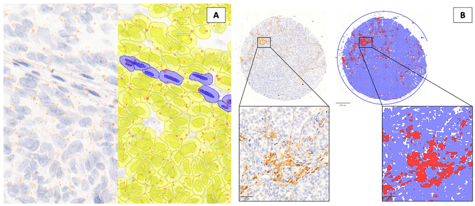
Figure 1 Detection of RNA hybridization signal and melanin deposits in melanoma formalin-fixed and paraffin-embedded tissues by digital image analysis. A) Subcellular detection of PPIB signal (orange dots) in tumor (yellow cells) and stromal (blue cells) compartments. B) Detection of melanin deposits by positive pixel count (red pixels = melanin; blue pixels = negative/no signal)
Serena Bonin and Eros Azzalini, University of Trieste.