
Digital pathology is an emerging sub-division of conventional microscopy, enabling practitioners to virtualize glass pathology slides for more in-depth analysis.
Artificial intelligence algorithms can assist pathologists with:
- Image analysis and interpretation
- Detailed inspections of sample tissues
- Pathology types matching to earlier cases
- Diagnosis accuracy and early detection
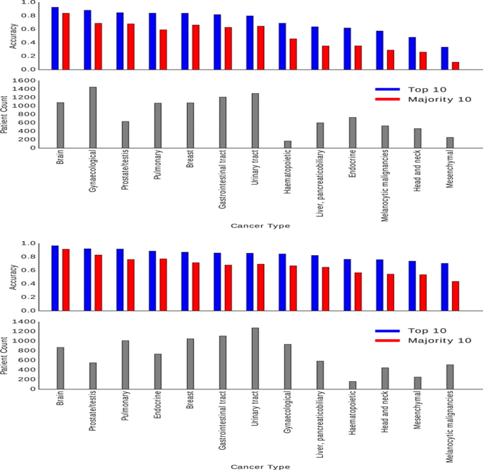
A group of cancer researchers recently analyzed a public database of WSIs from 11,000 cancer patients, featuring 32 cancer subtypes. The trained algorithm leveraged image data and annotations to reach a “computational consensus” on the type of pathology on display. The tool identified different types of pathology on frozen section slides with high accuracy:
- 93% for bladder urothelial carcinoma
- 97% for kidney renal clear cell carcinoma
- 99% for ovarian serous cystadenocarcinoma
And performed equally accurately with histopathology slides:
- 98% for prostate adenocarcinoma
- 99% for skin cutaneous melanoma
- 100% for thymoma
Similar artificial intelligence tools can support clinical decision-making and enhance the diagnosis of lesser studied pathologies and early-stage variations.