In the iToBoS project we will follow two slightly different data flows depending on the point of origin of the patient’s data, due to differences between data protection regulation in Europe vs. Australia.
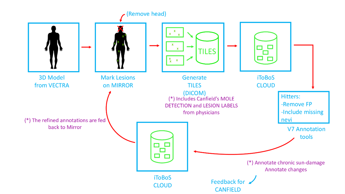
The data coming from the Clinical hospital from Barcelona will be collected by the VECTRA WB360 scanner (by Canfield), and this will provide a 3D model of the patient which includes skin texture. On that 3D avatar, the doctors annotate and classify the lesions of interest in order to facilitate their follow-up. The texture of this model is created from the 92 color images captured with the scanner, and the next step will consist on the anonymization of those images in order to store and share them among partners. To do that, Canfield has prepared a software (named MIRROR) that firstly detects the head automatically and removes it from each image so that the face of the patient is never visible. A similar operation is carried out if the patient has any tattoo, which could also lead to the recognition of the patient. The software also uses AI to perform a first detection of lesions, to reduce the work of annotators in following steps. Once this process is completed, the images are divided into several tiles so that the area of each tile is quite small (in the order of 8x8cm), thus anonymizing the patient’s skin images. Finally, the set of tiles are exported from the original images including the annotations that the doctors provided and excluding the texture on the anonymized areas. This data will then be converted into a DICOM file, packing both the color image and the annotation mask together in a single file, using different frame slots. At this point, the image files are completely anonymized and can be safely uploaded to the iToBoS Cloud.
In a second stage, the automatic lesion detections from the AI are then reviewed and cleaned by ISAHIT annotators, in order to ensure a good quality ground truth for the machine learning algorithms that will use these data as ground truth. Therefore, the tiles are uploaded on V7 annotation platform, where the “hitters” (ISAHIT annotators) will be able to delete all false positive automatic detections, and highlight any pigmented skin lesions that may not have been previously annotated by doctors (those correspond to darker lesions with a diameter of more than 3mm). If new lesions are included by the hitters, the doctors will be able to review those in the following steps. Every scan provides around 700 tiles, so this step is essential in order to be able to filter the tiles with suspicious lesions and significantly reduce the work performed by the doctors. Those corrected annotations will then be exported back to the cloud, and the doctors will review all of the suspicious lesions detected by the annotators, and classify each of them. This accurate ground truth will then be send back to the software running at the hospital in order to re-generate the tiles with the new ground truth, closing the loop that allows the system to be permanently refined.